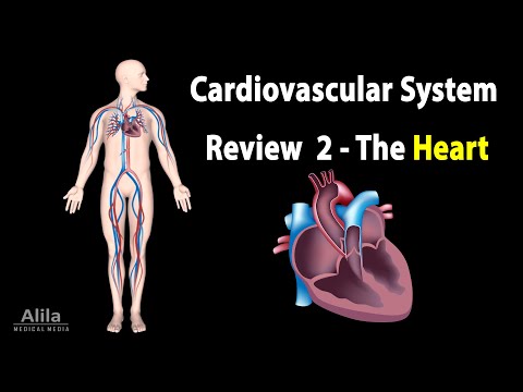
CARDIOVASCULAR SYSTEM REVIEW 2 – ALL ABOUT HEART in less than 10 min, Animation
– Oxygen-poor blood from the body returns to the right side of the heart, where it is pumped to the lungs. In the lungs, blood picks up oxygen and releases carbon dioxide. Oxygen-rich blood returns to the heart’s left side to be pumped to body’s tissues, where it unloads oxygen and picks up carbon dioxide. The resulting deoxygenated blood again returns to the heart’s right side to complete the cycle. – The 4 heart valves ensure one-way blood flow through the heart: oxygen-poor blood flows from the right atrium to right ventricle to pulmonary arteries; oxygen-rich blood moves from the
left atrium to left ventricle to the aorta. – The heart is enclosed in a double-layered sac called pericardium. The pericardial cavity contains a lubricant fluid. – The heart wall has 3 layers: epicardium, myocardium, and endocardium. Myocardium is the muscle tissue responsible for the beating of the heart. – The heart supplies itself via a network of blood vessels called the coronary circulation, which branches out from the aorta as soon as it exits the heart. A blockage in any major coronary artery may cause heart attacks. ——————————————- – The heart is essentially a muscle that contracts and pumps
blood. It consists of specialized muscle cells called cardiac myocytes. Contraction of these cells is initiated by electrical impulses, called action potentials. Unlike skeletal muscles,
and the signals spread rapidly. – Action potentials propagate through the heart in a sequence that follows the cardiac conduction pathway. The impulses start in the SA node, the heart’s primary pacemaker, then spread though the atria to reach the AV node. The AV node passes the signals onto AV bundle, then bundle branches, Purkinje fibers, and finally the ventricular myocytes. – Cells are polarized, they have different electric charges across their membrane. A resting cell has a negative membrane potential. When membrane potential increases and becomes less negative, the cell is less polarized, or depolarized. Reversely, when membrane voltage
becomes more negative, the cell is repolarized. Depolarization is due to net influx of positive ions. Repolarization is due to net efflux of positive ions. For an action potential to be generated, the membrane voltage must depolarize to a certain threshold value. – Pacemaker cells of the SA node fire 60 to 100 action potentials per minute. They do not have a true resting potential. Instead, “funny” channels, present only in pacemaker cells, open spontaneously when membrane voltage becomes lower than -40mV and allow influx of sodium, which brings the voltage back up to the threshold of -40mV. This is
“pacemaker potential”. Further depolarization is due to calcium influx. Repolarization is due to potassium efflux. – Contractile myocytes contain myofibrils, and a large amount of calcium stored in the sarcoplasmic reticulum, the SR. They have a stable resting potential and depolarize only when stimulated by a neighboring myocyte. – The depolarizing phase is due to fast sodium channels and slow calcium channels. Early repolarization is due to potassium efflux. Calcium channels, however, remain open and potassium efflux is eventually balanced by calcium influx, keeping the membrane potential relatively stable for about 200 milliseconds, resulting in the plateau phase. – Calcium
influx triggers a much greater calcium release from the SR, a process called “calcium-induced calcium release”. Calcium then sets off muscle contraction by the “sliding filament mechanism”. – Repolarization is due to potassium efflux with calcium channels closed. – Because of the plateau phase, cardiac muscle stays contracted longer than skeletal muscle. This is necessary for expulsion of blood from heart chambers. The absolute refractory period is also much longer to make sure the muscle has relaxed before it can respond to a new stimulus. This is essential to prevent summation and tetanus, which would stop the heart from beating.
———————————————- – While action potential waveform reflects electrical activities in one single cell, an ECG is a composite recording of all action potentials produced by the cells of the heart. ECG reflects electrical activities of the heart as a whole. – P wave represents the sum of depolarization in all atrial myocytes. – QRS complex reflects ventricular depolarization. Atrial repolarization also occurs during this time. – ST segment reflects the plateau phase of action potentials in ventricular myocytes. This is when the ventricles contract and pump blood. – T wave corresponds to ventricular repolarization that occurs immediately before ventricular relaxation.
———————————————— – A cardiac cycle has 2 major phases: systole, or ventricular contraction; and diastole, or ventricular relaxation; each with several smaller phases. – The following rules govern the events of the cardiac cycle: – Blood flows from higher to lower pressure; – Contraction increases the pressure within a chamber, while relaxation lowers its pressure; – AV valves open when atrial pressures are higher than ventricular pressures, and close when the pressure gradient is reversed. Semilunar valves operate similarly. – The cycle is initiated with the firing of the SA node that stimulates the atria to depolarize. Phase 1 includes
atrial depolarization and atrial contraction. – The closing of AV valves produces the first heart sound, S1, and marks the beginning of systole. The first part of systole, phase 2, is isovolumetric contraction, because the ventricles start to contract, but semilunar valves remain closed, and no blood is ejected. – Phase 3, rapid ejection, starts when ventricular pressures exceed the pressures within the aorta and pulmonary artery, pushing semilunar valves to open. – Phase 4 includes ventricular repolarization, and reduced ejection. – Closure of semilunar valves produces the second heart sound, S2, and marks the beginning of diastole. The first
part of diastole, phase 5, is again, isovolumetric, as the ventricles relax with all valves closed. – Phase 6, ventricular filling, starts when ventricular pressures drop below atrial pressures, causing AV valves to open. —————————————— – Cardiac output is the amount of blood pumped by each ventricle in one minute. It is the product of stroke volume – the amount of blood pumped in one heartbeat, and heart rate – the number of beats in one minute. – The ventricles do not eject all the blood they contain in one beat. The volume of blood in a ventricle at the
end of its filling is the end-diastolic volume, EDV. The volume that remains after contraction is the end-systolic volume, ESV. The percentage of blood ejected is the ejection fraction. Stroke volume equals EDV minus ESV, and is dependent on 3 factors: contractility, preload, and afterload. – Contractility is the force of contraction of the heart muscle. – Preload is the degree of stretch of cardiac myocytes at the end of ventricular filling. Preload is related to EDV. – Afterload is the resistance that the ventricle must overcome to eject blood. Afterload increases with vascular pressure and valve damage.